
Handheld Ultrasound The Next Stethoscope?
Will ges handheld ultrasound become the next stethoscope – Will handheld ultrasound become the next stethoscope? This introduction explores the potential of this revolutionary technology, examining its capabilities, limitations, and impact on healthcare. From its portability to its diagnostic power, we’ll dive into the comparison between this innovative tool and the traditional stethoscope, evaluating their respective roles in modern medicine.
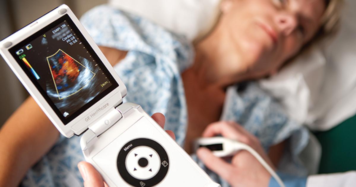
Handheld ultrasound technology has rapidly evolved, offering a detailed look at internal structures. This innovative tool provides real-time images, allowing for quick and accurate diagnoses, especially when compared to the limited visual perspective of a stethoscope. The potential benefits for healthcare providers and patients are substantial.
Introduction to Handheld Ultrasound: Will Ges Handheld Ultrasound Become The Next Stethoscope
Handheld ultrasound technology has rapidly evolved, transforming medical diagnostics and treatment. Its portability, combined with real-time imaging capabilities, offers advantages over traditional methods, particularly in point-of-care settings. This technology has revolutionized various fields, from emergency medicine to obstetrics, allowing for quick and accurate assessments.The core concept behind handheld ultrasound involves emitting high-frequency sound waves that bounce off internal structures.
The returning echoes are processed to create images, providing clinicians with valuable information about the body’s anatomy and physiology. This capability is dramatically changing how medical professionals approach patient care, enabling immediate assessments and potentially faster interventions.
Key Features and Applications, Will ges handheld ultrasound become the next stethoscope
Handheld ultrasound devices offer a range of features that contribute to their increasing popularity. These devices are lightweight and compact, making them easily transportable to various locations. Real-time imaging provides immediate visual feedback, allowing for dynamic assessments of organs and tissues. This immediacy is crucial in emergency situations where rapid diagnosis and intervention are critical. Applications span a broad spectrum, from assessing soft tissue injuries in sports medicine to guiding biopsies in interventional procedures.
Additionally, handheld ultrasound is used in obstetrics for fetal monitoring and in vascular assessments for detecting blockages.
Evolution of Handheld Ultrasound Devices
Early handheld ultrasound devices were often bulky and limited in their functionality. Technological advancements have led to significant improvements in size, weight, image quality, and processing speed. These improvements have made the technology more accessible and practical for diverse clinical settings. The miniaturization and integration of advanced imaging algorithms have allowed for higher resolution and greater detail in the images.
This evolution is directly comparable to the development of smartphones; the initial devices were large and limited in capabilities, but subsequent iterations have become smaller, more powerful, and more integrated into our daily lives.
Market Penetration and Adoption Rates
The adoption rate of handheld ultrasound devices is steadily increasing. Its integration into various medical specialties, particularly emergency medicine and point-of-care settings, has contributed to this growth. The increasing availability of affordable, high-quality devices is driving wider adoption. Examples include emergency rooms, urgent care centers, and even remote clinics, showcasing a shift towards more accessible diagnostic tools.
Advantages and Disadvantages Compared to Traditional Methods
Handheld ultrasound offers several advantages over traditional diagnostic methods like X-rays or CT scans. Its portability allows for point-of-care imaging, reducing the need for patients to travel to specialized facilities. Real-time imaging provides immediate feedback, aiding in rapid diagnosis and intervention. Furthermore, handheld ultrasound is often less expensive than traditional imaging modalities, making it a more cost-effective solution in certain situations.
Will handheld ultrasound devices from companies like GE become the next stethoscope? It’s a fascinating question, especially with the recent news that Google is testing new technologies with early adopters, like in their google waves hello to early testers program. Perhaps the advancements in portable tech, similar to Google’s innovations, will pave the way for these ultrasounds to become more accessible and commonplace, eventually making them as ubiquitous as stethoscopes are today.
However, handheld ultrasound may have limitations in visualizing certain structures compared to more sophisticated imaging techniques. The quality of images can sometimes be affected by patient positioning or body habitus. In certain cases, traditional methods may be required for more comprehensive assessments.
Comparison to Stethoscopes
| Feature | Handheld Ultrasound | Stethoscope |
|---|---|---|
| Portability | High – easily transported and used in various settings | High – compact and lightweight |
| Cost | Variable – depends on the device and features | Low – generally inexpensive |
| Image Quality | High – visualizes internal structures | Low – auditory assessment, limited anatomical detail |
| Accessibility | Increasing – expanding use in various specialties | High – widely available and easy to use |
The table above highlights the key differences between handheld ultrasound and stethoscopes, emphasizing their distinct capabilities and applications. Stethoscopes remain valuable for auscultation, but handheld ultrasound offers the advantage of visualizing internal structures in real time. This visualization capability is invaluable for diagnosis and intervention in various medical specialties.
Comparison with Stethoscopes
Handheld ultrasound technology is rapidly transforming healthcare, offering a powerful tool for diagnosing a wide range of medical conditions. While stethoscopes remain fundamental diagnostic instruments, their limitations in visualizing internal structures are increasingly apparent. This comparison highlights the superior capabilities of handheld ultrasound, especially in situations demanding a more comprehensive view of the patient’s anatomy.The stethoscope primarily relies on detecting sound waves produced by internal organs.
Its diagnostic power is limited to identifying abnormalities in sound characteristics, such as altered heart murmurs or lung sounds. Handheld ultrasound, however, provides a visual representation of the structures and tissues, enabling a deeper understanding of the underlying anatomy and pathology.
Will handheld ultrasound devices from GES become the next stethoscope? It’s a fascinating question, especially considering the recent Nokia Expo, which unveiled some exciting new phone features and Facebook updates. Nokia expo ushers in fresh phones facebook features might just hint at a future where medical diagnostics are more accessible and integrated with our daily tech lives.
Perhaps these handheld ultrasound tools, with their potential for on-the-spot diagnoses, will eventually replace the traditional stethoscope as the primary diagnostic tool in many settings.
Diagnostic Capabilities
Stethoscopes are effective for assessing general cardiovascular and respiratory function, detecting abnormal heart murmurs, and identifying wheezing or crackles in the lungs. However, their ability to identify subtle structural changes, or conditions affecting deeper tissues and organs, is significantly limited. For example, a subtle mass in the abdomen, a fluid collection in the lungs, or a blood clot in a vessel cannot be visualized with a stethoscope.
Limitations of Stethoscopes
Stethoscopes are limited by their inability to visualize internal structures. They rely on detecting subtle changes in sound patterns, which can be misleading or inconclusive in certain situations. Conditions like a thickening of the heart muscle, a cyst in the kidney, or a tumor in the breast may not manifest in audible cues detectable by a stethoscope. This limitation restricts their diagnostic utility for a range of conditions that require detailed structural evaluation.
Benefits of Ultrasound
Handheld ultrasound, unlike stethoscopes, provides real-time, high-resolution images of internal structures. This visual data offers a far more comprehensive understanding of the underlying pathology, enabling clinicians to identify a broader spectrum of conditions. For example, in evaluating abdominal pain, ultrasound can quickly differentiate between a simple gas buildup, a gallstone, or a significant obstruction. Similarly, in cases of suspected musculoskeletal injuries, ultrasound can identify tears in tendons or ligaments, offering a non-invasive alternative to more invasive procedures.
Situations Favoring Ultrasound
Handheld ultrasound becomes the preferred diagnostic tool in situations requiring visualization of internal structures. This includes, but is not limited to, evaluating abdominal masses, assessing the presence of fluid collections in body cavities, or evaluating the integrity of musculoskeletal structures. When precise anatomical details are crucial for accurate diagnosis, ultrasound provides superior insight compared to a stethoscope.
Comparison Table
| Medical Condition | Stethoscope | Handheld Ultrasound |
|---|---|---|
| Heart Murmurs | Effective | Useful for evaluating underlying cardiac structures |
| Lung Sounds | Effective | Essential for evaluating lung tissues and fluid |
| Abdominal Pain | Limited in identifying the source | Essential for differentiating between various causes (e.g., gas, gallstones, obstruction) |
| Musculoskeletal Injuries | Limited in identifying soft tissue damage | Provides detailed images of tendons, ligaments, and muscles |
| Suspected Pregnancy | Limited | Useful for confirming intrauterine pregnancy and evaluating fetal health |
| Kidney Cysts | Unlikely to detect | Visualizes the cyst, its size, and location |
Potential Market Impact

Handheld ultrasound, with its growing accessibility and affordability, has the potential to revolutionize the way healthcare professionals diagnose and treat patients. Its ability to provide real-time, non-invasive anatomical visualization could significantly impact various medical specialties, leading to earlier diagnoses, more accurate treatment plans, and improved patient outcomes. This shift from relying solely on stethoscopes to integrating ultrasound into routine practice will necessitate adjustments across the entire healthcare landscape.The adoption of handheld ultrasound as a routine diagnostic tool will dramatically alter healthcare providers’ workflows.
Instead of relying solely on auscultation, physicians, nurses, and other healthcare professionals will be able to visualize internal structures, enabling quicker assessments of conditions like fluid buildup, organ size, and tissue integrity. This visual confirmation can lead to faster, more informed decisions, potentially reducing diagnostic delays and improving patient care.
Impact on Healthcare Professionals’ Workflow
The integration of handheld ultrasound into daily practice will require healthcare professionals to adapt their workflows. Instead of just listening for sounds, they will be looking at images. This will involve a learning curve, but the benefits are substantial. For example, a physician examining a patient with suspected appendicitis might use ultrasound to quickly visualize the appendix and surrounding tissues, confirming the diagnosis or ruling it out.
Similarly, a nurse monitoring a patient’s fluid balance could use ultrasound to evaluate the amount of fluid in the abdomen, helping adjust treatment strategies.
Impact on Medical Training and Education
Medical training programs will need to adapt to incorporate handheld ultrasound into their curricula. This will involve incorporating new skills, from basic operation of the device to interpretation of images. New educational resources, workshops, and continuing medical education programs will be essential to ensure healthcare professionals are adequately trained and proficient in using the technology. Specialized training for specific medical specialties, such as cardiology or obstetrics, will also be necessary.
Impact on Healthcare Costs and Accessibility
The potential cost savings from using handheld ultrasound could be significant. By enabling earlier and more accurate diagnoses, it can potentially reduce the need for expensive and invasive procedures. For example, avoiding unnecessary exploratory surgeries based on ultrasound findings could result in substantial cost savings. Furthermore, wider adoption could increase accessibility to diagnostic tools in underserved areas, where traditional imaging may not be readily available.
This improved accessibility is crucial for improving health outcomes, especially in rural or resource-constrained environments.
Potential Training Needs
| Skill Area | Description | Training Duration (Estimated) |
|---|---|---|
| Basic Ultrasound Operation | Learning to operate the device, including positioning, image optimization, and basic controls. | 1-2 days |
| Image Interpretation | Understanding the various anatomical structures and recognizing normal and abnormal findings. | 1-3 weeks (depending on specialty) |
| Specific Application (e.g., Cardiac Ultrasound) | Learning to perform and interpret specialized ultrasound examinations. | 2-6 weeks (depending on specialty and complexity) |
| Patient Safety and Infection Control | Adhering to safety protocols and infection control measures during procedures. | Ongoing training |
This table Artikels potential training needs for healthcare professionals seeking to integrate handheld ultrasound into their practice. The specific duration of training will vary based on the individual’s prior experience and the complexity of the intended applications.
Barriers to Adoption

Handheld ultrasound technology, while promising, faces significant hurdles to widespread adoption as the next stethoscope. These challenges, spanning cost, training, accessibility, and infrastructure, require careful consideration to fully realize the potential of this technology in various healthcare settings. Overcoming these barriers will be crucial for ensuring equitable access and integration into existing medical practices.
Cost Considerations
The initial investment for handheld ultrasound devices, while decreasing, can still be substantial for individual clinics or smaller healthcare facilities. The cost includes the device itself, accessories like probes, and potential ongoing maintenance and software updates. This financial burden could limit access, particularly in resource-constrained environments. Furthermore, the cost of training personnel to operate and maintain the equipment must be factored in.
The overall cost-benefit ratio needs to be carefully assessed for different healthcare settings.
Training Requirements
Adequate training is essential for effective use of handheld ultrasound. Clinicians need training on image interpretation, proper probe technique, and the specific applications relevant to their practice. Training programs must be tailored to various healthcare settings and skill levels. This requires investment in training resources, qualified instructors, and potentially specialized training centers. Without proper training, the technology’s potential benefits will be limited, leading to suboptimal outcomes and potentially inaccurate diagnoses.
Accessibility and Infrastructure
Accessibility varies significantly across different healthcare settings. In remote areas, logistical challenges, limited infrastructure, and the need for specialized maintenance can hinder the integration of handheld ultrasound. Hospitals and clinics also face logistical issues in integrating new technology into existing workflows. Ensuring the availability of power, sufficient space, and appropriate storage for the devices is critical. In addition, the need for reliable internet connectivity for image transmission and software updates must be addressed.
Regulatory Hurdles
The adoption of handheld ultrasound also faces regulatory hurdles. Regulatory approval processes can vary significantly by region and may involve clinical trials, safety assessments, and compliance with specific standards. These processes can be lengthy and complex, further delaying widespread adoption. Potential issues related to liability, data security, and image interpretation accuracy must be addressed in the regulatory framework.
Challenges Across Healthcare Settings
| Healthcare Setting | Challenges |
|---|---|
| Clinics | Limited space and budget for initial investment; integration with existing workflows; training personnel in diverse specialties; ongoing maintenance needs. |
| Hospitals | Integration with existing imaging departments; potential workflow conflicts; need for dedicated training spaces and personnel; managing potential overload on imaging resources. |
| Remote Areas | Transportation and logistical issues for device maintenance; limited internet connectivity; difficulty in obtaining trained personnel; availability of power sources; potential for device malfunction in harsh environments. |
Future Trends and Projections
Handheld ultrasound technology is rapidly evolving, promising to reshape the landscape of medical diagnostics and treatment. Its accessibility, portability, and cost-effectiveness are driving significant advancements, leading to broader applications across various healthcare settings. This evolution is not just about incremental improvements; it’s about a fundamental shift in how we approach medical imaging and patient care.The future of handheld ultrasound is not confined to incremental improvements; it’s about a paradigm shift in how we approach medical imaging and patient care.
We can anticipate a future where these devices become even more integrated into daily clinical practice, offering real-time insights and empowering healthcare professionals with crucial diagnostic tools.
Potential Future Development of Handheld Ultrasound Technology
Advancements in sensor technology, particularly in image processing and data analysis, will be pivotal. Improved image resolution and contrast will allow for more detailed visualization of internal structures, leading to more accurate diagnoses. Real-time 3D imaging capabilities, now being researched, will provide a more comprehensive understanding of anatomical structures and potentially reduce the need for more invasive procedures.
Will handheld ultrasound devices from GE become the next generation stethoscope? It’s a fascinating question, and the answer might lie in the future of medical diagnostics. This technology has the potential to revolutionize how we approach patient care, offering a deeper look at the body than ever before. However, the complexities of integrating such technology with current medical practice and the questions of cost-effectiveness need careful consideration.
The article whos afraid of the big bad wolf huntress offers some interesting insights into the future of technology in medicine, and the potential of similar innovations. Ultimately, whether or not GE’s handheld ultrasound devices truly become the next stethoscope remains to be seen, but the possibilities are certainly exciting.
Emerging Technologies Enhancing Functionality
Several emerging technologies are poised to enhance the functionality of handheld ultrasound devices. Artificial intelligence (AI) is poised to play a significant role. AI algorithms can be trained to analyze ultrasound images, potentially identifying subtle anomalies that might be missed by human operators. This could lead to earlier detection of diseases and improved diagnostic accuracy. Moreover, integration with wearable sensors and patient monitoring devices will provide valuable contextual information, enabling clinicians to make more informed decisions.
The development of new, more compact and user-friendly interfaces will further increase accessibility.
Integration with Other Medical Devices and Technologies
The potential for integration with other medical devices is substantial. Imagine a future where a handheld ultrasound device could seamlessly connect to an electronic health record (EHR) system, instantly transmitting images and reports. Integration with telemedicine platforms could allow remote diagnoses and consultations, especially in underserved areas. Further, integration with robotic surgical systems could provide real-time guidance during minimally invasive procedures, leading to more precise and less traumatic interventions.
Potential Future Applications in Diverse Healthcare Fields
The applications of handheld ultrasound extend beyond traditional uses in cardiology and obstetrics. In emergency medicine, it can provide rapid assessments of trauma patients, facilitating quicker interventions. In primary care, it can be used for routine screenings and monitoring of chronic conditions. Furthermore, it’s anticipated that handheld ultrasound will play a critical role in point-of-care diagnostics, potentially revolutionizing the way healthcare is delivered in resource-limited settings.
Table of Potential Future Features and Advancements
| Feature | Description | Impact |
|---|---|---|
| AI-Powered Image Analysis | Integration of AI algorithms for automatic image analysis and anomaly detection. | Increased diagnostic accuracy, reduced human error, and potentially faster diagnoses. |
| 3D/4D Imaging Capabilities | Real-time 3D/4D imaging to provide more comprehensive anatomical visualization. | Enhanced understanding of anatomical structures and potential reduction in invasive procedures. |
| Integration with Wearable Sensors | Connecting to patient monitoring devices to provide contextual information. | Improved patient monitoring and more informed decision-making. |
| Wireless Connectivity and Cloud Integration | Seamless transmission of data and images to EHRs and remote clinicians. | Improved efficiency, accessibility, and communication among healthcare providers. |
| Miniaturization and Portability | More compact and lightweight devices for improved portability and accessibility. | Wider applications in diverse healthcare settings, including remote areas. |
Illustrative Case Studies
Handheld ultrasound is rapidly gaining traction in various medical settings, offering a valuable alternative to traditional diagnostic methods. Its portability, real-time imaging capabilities, and relative affordability make it a promising tool for improving patient care, particularly in resource-constrained environments. These case studies illustrate how handheld ultrasound can enhance diagnostic accuracy and efficiency, supplementing or even replacing traditional methods in specific clinical scenarios.This section presents hypothetical case studies demonstrating the advantages of handheld ultrasound, focusing on scenarios where it provides superior or complementary information compared to a stethoscope alone.
Each example highlights the specific benefits of the technology, including rapid assessment, visualization of structures, and identification of subtle abnormalities that might be missed with auscultation alone.
Case Study 1: Suspected Appendicitis
A 16-year-old patient presents with right lower quadrant pain, fever, and nausea, suggestive of appendicitis. A preliminary physical exam, including auscultation, reveals tenderness but no specific, definitive findings. Handheld ultrasound, however, can directly visualize the appendix and surrounding structures. It can determine the presence of appendicitis by identifying an inflamed or enlarged appendix, as well as signs of possible complications such as abscess formation.
This visual confirmation can significantly aid in the diagnostic process and potentially expedite treatment decisions. In cases of uncertainty, the ability to quickly and accurately visualize the appendix through handheld ultrasound can be invaluable in guiding further treatment strategies, such as surgical intervention.
Case Study 2: Evaluation of a Distended Abdomen
A 75-year-old patient with a history of congestive heart failure presents with a significantly distended abdomen. While a stethoscope might reveal fluid build-up, a handheld ultrasound can differentiate between ascites (fluid in the peritoneal cavity), and other conditions like masses or gas. The real-time imaging capabilities of handheld ultrasound allow clinicians to evaluate the extent and distribution of fluid, providing crucial information for treatment planning.
Differentiation is critical as treatment strategies vary depending on the underlying cause. This case illustrates the value of ultrasound in providing a clearer picture of the cause of the distension, guiding treatment decisions more effectively.
Case Study 3: Assessing a Post-Surgical Patient
A patient undergoing a recent surgical procedure complains of persistent pain in the surgical site. A stethoscope may reveal some signs, but a handheld ultrasound can provide a deeper understanding of the surgical site. It can visualize any collections of fluid, hematomas, or other complications that might not be apparent through auscultation alone. This real-time assessment allows clinicians to make more informed decisions regarding pain management and potential interventions, facilitating quicker recovery.
Early detection of complications, like postoperative hematomas, is vital to prevent further complications and ensure patient well-being.
Comparison of Diagnostic Approaches: Stethoscope vs. Handheld Ultrasound
| Diagnostic Tool | Scenario | Diagnostic Findings | Limitations |
|---|---|---|---|
| Stethoscope | Assessing for possible pneumonia | Can detect crackles and wheezes, suggesting inflammation. | Can’t visualize the lungs or assess for fluid buildup; may not distinguish between different types of lung issues. |
| Handheld Ultrasound | Assessing for possible pneumonia | Can visualize lung structures, identify consolidation, pleural effusion, or other abnormalities. | Requires specialized training to interpret images; might not be readily available in all clinical settings. |
This table highlights the comparative strengths and weaknesses of both methods. In cases of suspected pneumonia, a stethoscope might suggest inflammation, but handheld ultrasound provides a visual confirmation of the underlying cause and severity, offering a more comprehensive evaluation. This crucial visualization can affect the treatment strategy.
Summary
Ultimately, the question of whether handheld ultrasound will replace the stethoscope entirely is complex. While ultrasound offers superior diagnostic capabilities in many cases, the stethoscope remains invaluable for certain assessments. The future likely lies in a complementary approach, where both tools are used strategically to provide the most comprehensive and effective patient care.